World Journal of Nephrology and Urology, ISSN 1927-1239 print, 1927-1247 online, Open Access
Article copyright, the authors; Journal compilation copyright, World J Nephrol Urol and Elmer Press Inc
Journal website https://www.wjnu.org

Case Report

Volume 12, Number 1, July 2023, pages 11-16


A Rare Case of Testicular Plasmacytoma as First Presentation in a Monorchid Patient Without Previous History of Multiple Myeloma: Diagnosis, Management and Review of Literature

Figures

Figure 1.
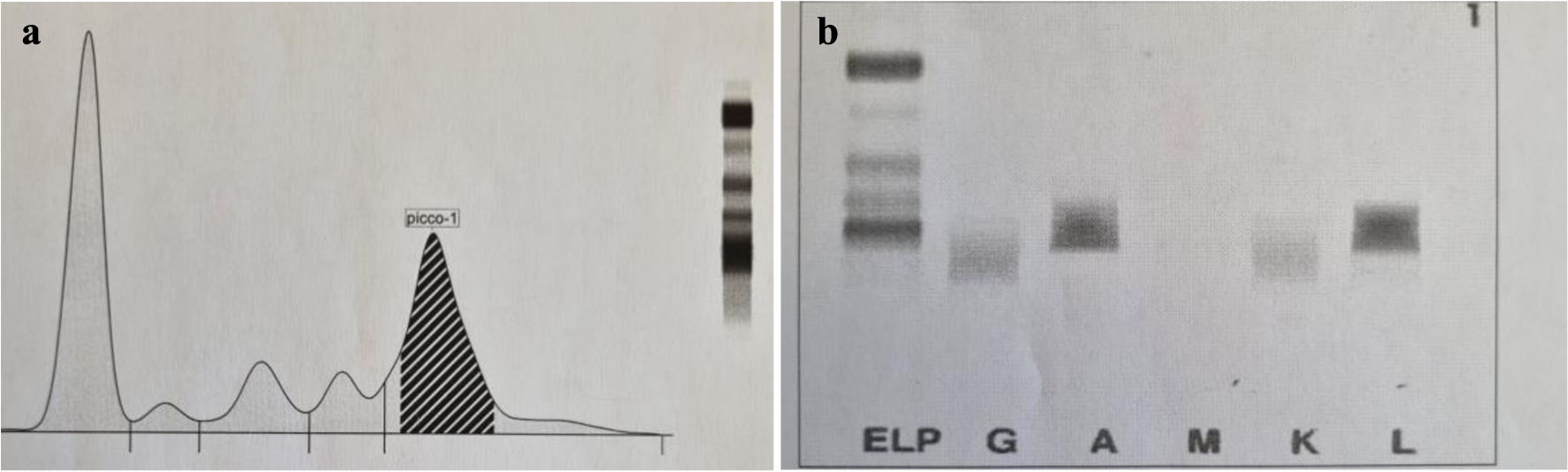
Figure 1. (a) Electrophoresis of serum protein showing gamma-spike. (b) Urine electrophoresis showing monoclonal light chains.
Figure 2.
Figure 2. On gross examination, testis showed neoformation sized 3.3 × 2.5 cm, variegated in appearance with necrotic areas.
Figure 3.
Figure 3. (a) Diffuse infiltration of atypical plasma cells surrounding atrophic seminiferous tubule (arrow) (hematoxylin and eosin; × 200). (b) A population of immature and anaplastic plasma cells predominated, with a higher nuclear/cytoplasmic ratio, scattered nuclear chromatin and often prominent nucleoli, with occasional intracellular small vacuoles (arrow) (hematoxylin and eosin; × 400).
Figure 4.
Figure 4. Neoplastic plasma cells showing immunohistochemical expression of CD38 at the membrane level (a) and nuclear staining of MUM1 (b) (× 200).
Figure 5.
Figure 5. (a) Immunohistochemical study confirming the monoclonality of neoplastic process by showing restriction to lambda light chains (a, × 200) and negativity to kappa chains (b ,× 170).