World Journal of Nephrology and Urology, ISSN 1927-1239 print, 1927-1247 online, Open Access
Article copyright, the authors; Journal compilation copyright, World J Nephrol Urol and Elmer Press Inc
Journal website http://www.wjnu.org

Case Report

Volume 4, Number 2, June 2015, pages 222-225


Beyond the Boundaries: Enigma of Distinguishing Exophytic Upper Pole Renal Cell Carcinoma From an Adrenal Mass

Figures

Figure 1.
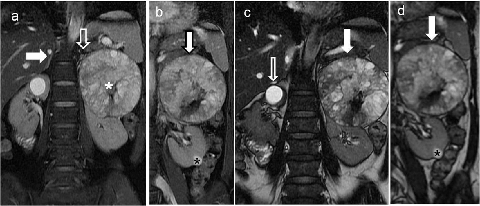
Figure 1. Non-contrast magnetic resonance imaging (MRI) of the upper abdomen showing left retroperitoneal soft tissue heterogenous renal/adrenal mass with simple cyst in the right kidney. (a) Coronal T2-weighted fat suppressed sequence of the upper abdomen showing well visualized right adrenal gland (block white arrow) and the small partially visualized left adrenal gland (white arrow) indistinct from the soft tissue mass (white asterisk) along the left renal upper pole. (b) Coronal T2-weighted fat suppressed sequence of the upper abdomen showing soft tissue mass (bold white arrow with black outline), appearing predominantly T2 hyperintense and abutting the upper left renal pole. A small T2 hyperintense lesion in the renal cortex is also seen at the lower pole of left kidney (black asterisk). (c) Coronal FIESTA sequence of the upper abdomen showing the soft tissue mass abutting the left renal upper pole (bold white arrow). In addition, a brighter well rounded simple cortical cyst is present in the right renal upper pole (bold black and white arrow). (d) Coronal FIESTA sequence of the upper abdomen showing a relatively less bright rounded lesion in the lower pole of the left kidney (asterisk) in comparison to the cortical cyst seen in right kidney in (c).
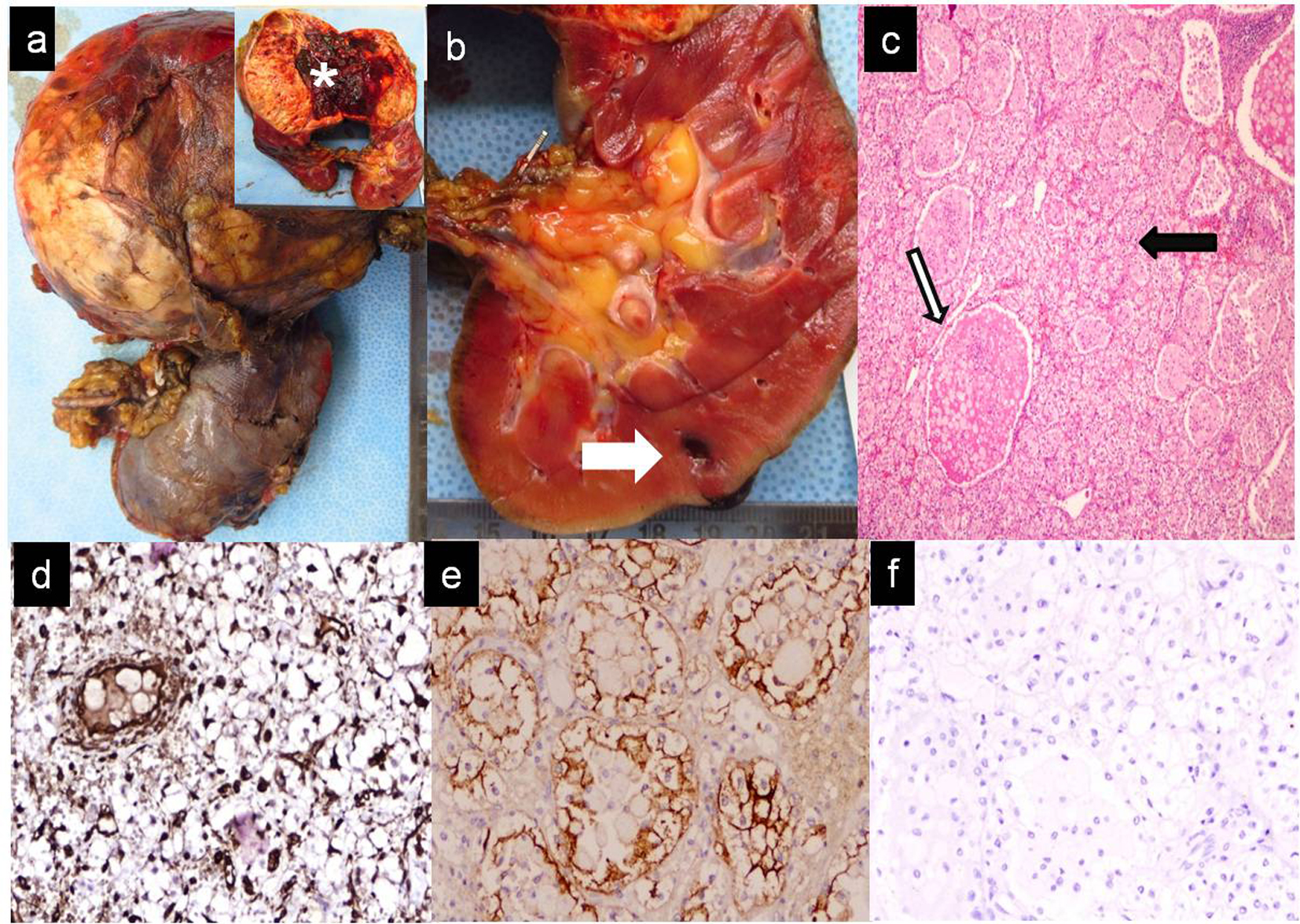
Figure 2.
Figure 2. Gross, microscopic and immunohistochemistry findings of the soft tissue suprarenal mass removed along with radical left nephrectomy. (a) On gross pathology examination, globular tumor weighed approximately 800 g, measured 11 × 9 × 8 cm. Cut surface (inset) partially hemorrhagic, solid-cystic (white asterisk) arising from the cortico-medullary junction of the left kidney. No involvement of pelvicalyceal system, renal vessels, lymphatics or of the left ureter. (b) Small intra-parenchymal cortical lesion (0.5 cm maximum dimension) in the lower pole (white bold arrow). (c) Microscopic examination of the tumor revealed a cellular tumor with nested, acinar and microcystic arrangement of malignant epithelial cells. Tumor cells displayed irregular enlarged nuclei with small nucleoli, clear cytoplasm, abundant cytoplasmic glycogen and a well-defined cell membrane (black arrow bold). These were interspersed within a highly vascularized stroma. Many glandular lumina showed presence of red blood cells (white bold arrow with black margins). (d-f) Immunohistochemistry revealed positive staining for vimentin (d), EMA (e) and negative staining for inhibin (f).

Tables

Table 1. Laboratory Investigations
 
Test performedResultsReference range
Hemoglobin13.012 - 15 g/dL
Platelets160150 - 400 × 109/L
TLC10.14 - 11 × 103/L
DLCN37L50M06E07N: 40-75%, L: 20-45%, M: 2-10%, E: 1-6%
Urea34.415 - 40 mg/dL
Cr0.440.2 - 1 mg/dL
Na/K144.4/3.7Na: 136 - 145, K: 3.5 - 5
Total bilirubin/direct bilirubin0.3/0.10.3 - 1.2 mg/dL/0.2 g/dL
AST/ALT21/225 - 40, 7 - 35 IU/L respectively
GGT87 - 64 IU/L
Total protein/Alb6.7/3.76 - 8/3.5 - 5.2 g/dL respectively
Urine routineUnremarkable
Urine cultureSterile
24-h urinary markers
  Metanephrines81.9127 - 155
  Nor-metanephrines168.7946 - 256
  VMA4.141.6 - 4.2
  Epinephrines3.571.3 - 10.7
  Nor-epinephrines15.368.9 - 61.60
  Dopamine185.1840 - 390


Table 2. Immunohistochemistry Markers to Differentiate RCC From Adrenal Cancer
 
IHC markersRCCACC
EMA+-
Vimentin+-
Carbonic anhydrase-IX+-
PAX-8+-
Calretinin-+
Inhibin-+
Melan A-+
Adrenal cortical antigens SF-1-+