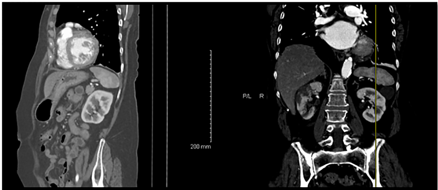
![Figure 2.](/tables/wjnu242e-g002.jpg)
Figure 2. A diagram of the most common pattern of arterial supply to the kidneys demonstrating the main renal artery, anterior and posterior branches, and five segmental arteries. MRA: main renal artery; PD: posterior division; AD: anterior division. Segmental arteries are indicated by A (apical), U (upper), M (middle), L (lower), and P (posterior). Modified from Graves FT. The anatomy of the intrarenal arteries and its application to segmental resection of the kidney. Br J Surg 1954;42:132.
![Figure 3.](/tables/wjnu242e-g003.jpg)
Figure 3. An allograft kidney from a 58-year-old obese man with a past medical history of hypertension and diabetes mellitus, who expired due to acute myocardial infarction and was found to have ARI on autopsy.
![Figure 5.](/tables/wjnu242e-g005.jpg)
Figure 5. Microscopic image of an arterial thrombus showing a vessel completely occluded by the thrombus (black arrow) and the resulting renal infarct (in and around circle) (from Fig. 4) (H&E, × 2.5).