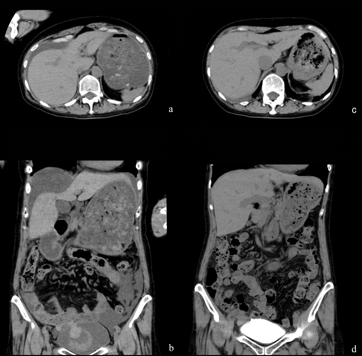
Figure 1. Findings from CT. (a) Non-contrast CT at the upper abdominal level showed a large amount of ascites at admission. (b) Non-contrast CT with coronal reconstruction at the urinary bladder level showed fresh blood clots in the bladder at admission. (c) Non-contrast CT at the upper abdominal level showed a small amount of ascites at 4 days after admission. (d) A CT cystogram with coronal reconstruction at the urinary bladder level showed no extravasation of contrast agent from the bladder dome at 14 days after admission.

