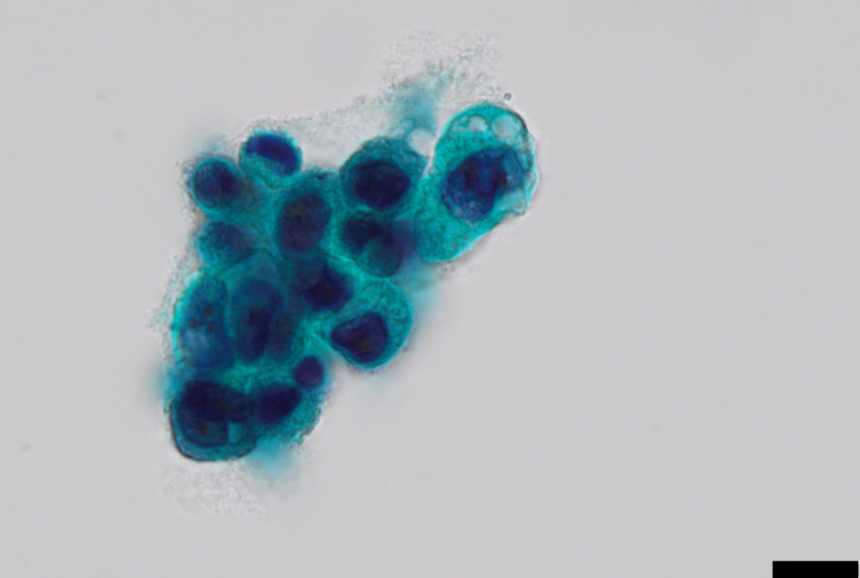
Figure 1. Urinary cytology showing a cluster of atypical urothelial cells with increased nuclear/cytoplastic ratio and grooved nuclei (Papanicolaou stain, scale bar = 10 µm).
| World Journal of Nephrology and Urology, ISSN 1927-1239 print, 1927-1247 online, Open Access |
| Article copyright, the authors; Journal compilation copyright, World J Nephrol Urol and Elmer Press Inc |
| Journal website https://www.wjnu.org |
Case Report
Volume 000, Number 000, September 2024, pages 000-000
Leiomyoma Associated With Epithelial Dysplasia of the Urinary Bladder: Cytopathologic Features and Review of the Literature
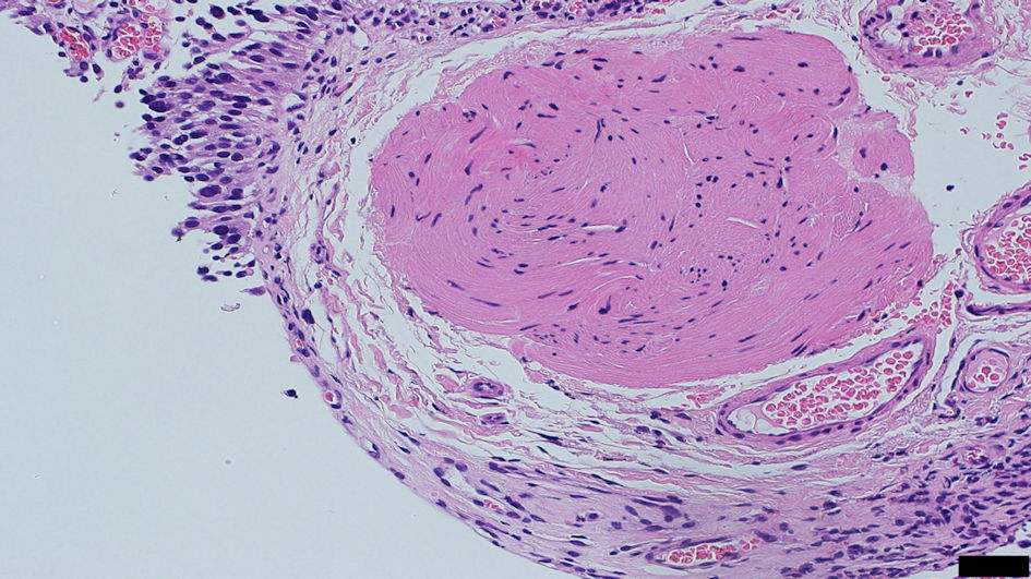
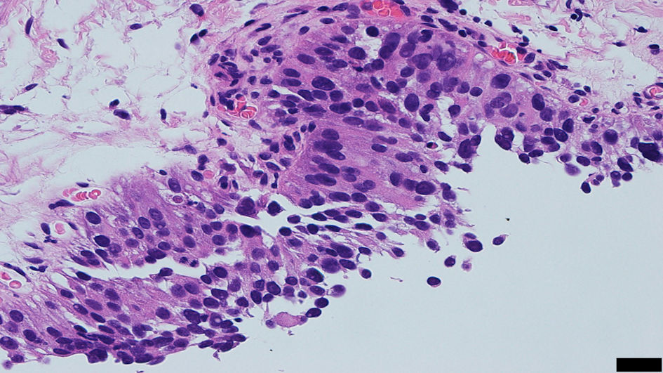
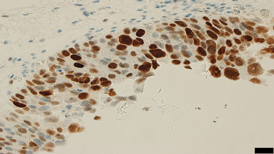
Figures