

| World Journal of Nephrology and Urology, ISSN 1927-1239 print, 1927-1247 online, Open Access |
| Article copyright, the authors; Journal compilation copyright, World J Nephrol Urol and Elmer Press Inc |
| Journal website http://www.wjnu.org |
Letter to the Editor
Volume 1, Number 2-3, June 2012, pages 98-98
Entrapment of the Left Renal Vein: A Rare Cause of Proteinuria
Sik Leea, c, Young Min Honga, Kyung Pyo Kanga, Won Kima, Sung Kwang Parka, Jong Kwan Parkb
aDepartment of Internal Medicine, Renal Research Institute of Clinical Medicine, Chonbuk National University Medical School, Jeonju, Republic of Korea
bDepartment of Urology, Renal Research Institute of Clinical Medicine, Chonbuk National University Medical School, Jeonju, Republic of Korea
cCorresponding author: Sik Lee, Department of Internal Medicine, Chonbuk National University Medical School, 634-18, Keum-Am Dong, Jeonju, 561-712, Republic of Korea
Manuscript accepted for publication June 11, 2012
Short title: Entrapment of the Left Renal Vein
doi: https://doi.org/10.4021/wjnu28w
| To the Editor | ▴Top |
A 26-year-old man visited our institution for evaluation of proteinuria. He intermittently presented with acute urinary retention. Other urological evaluations were unremarkable. Urinalysis exhibited trace proteins without hematuria. Renal duplex and enhanced computed tomography (CT) of the abdomen and pelvis were performed. The duplex ultrasound typically demonstrated aortomesenteric anatomy with entrapment of the left renal vein (LRV) (Fig. 1). The diameters of the LRV at the hilar and aortomesenteric portions were measaured as 11 mm and 1.2 mm, respectively. The duplex ultrasound also indicated a significantly higher ratio of the peak systolic velocity (PSV) to the hilar renal vein PSV at the point of renal vein compression (> 6). This observation demonstrates the diminished volume of flow and dampened pulsatility upon spectral analysis of the left renal vein. Enhanced CT angiography revealed compression of the left renal vein between the aorta and the superior mesenteric artery (Fig. 2). The Nutcracker syndrome is defined as a rare cause of hematuria and flank pain caused by compression of the left renal vein between the aorta and the superior mesenteric artery where it passes at the point of bifurcation of the arteries [1]. The observed compression causes venous hypertension and the formation of renal venous collaterals leading to the subsequent rupture of the septum between the veins and the collecting system, and finally, resulting in hematuria and flank pain [2, 3]. In this particular patient case, although rare, the nutcracker should be included as the potential cause of proteinuria in addition to hematuria in the absence of other renal diseases.
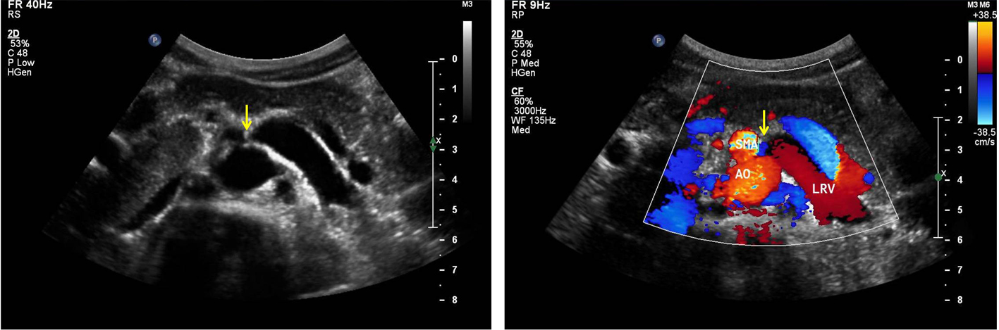 Click for large image | Figure 1. The duplex ultrasound typically demonstrated aortomesenteric anatomy with entrapment of the left renal vein (LRV). |
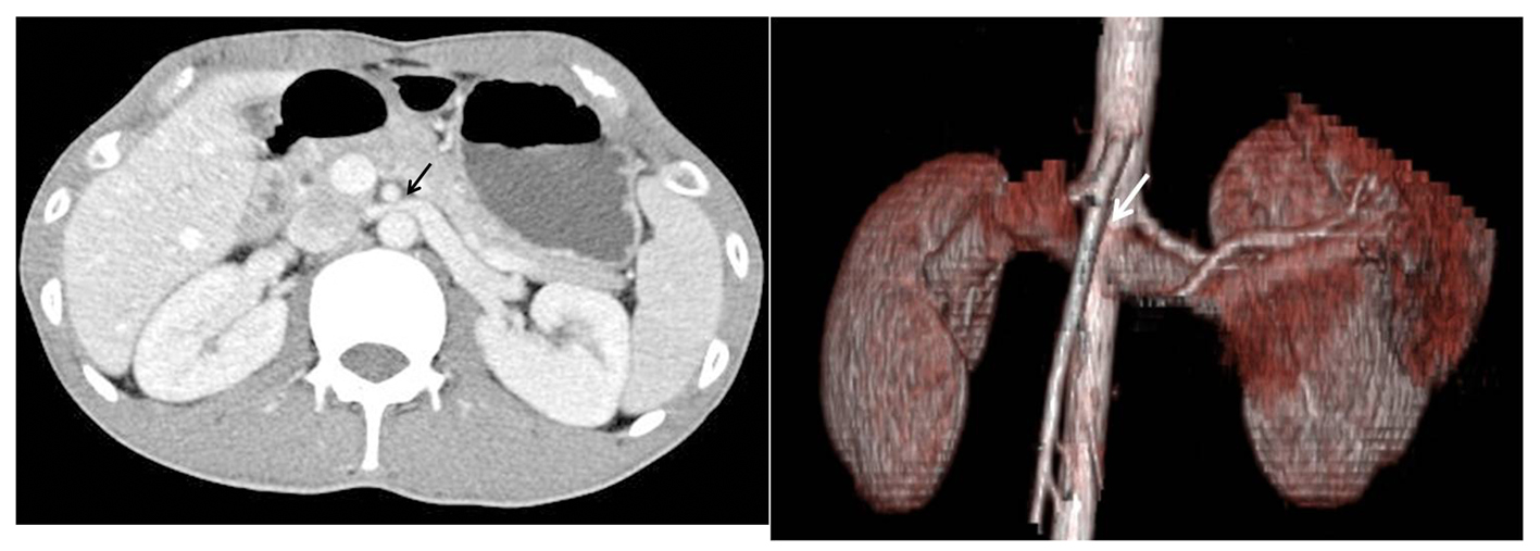 Click for large image | Figure 2. Enhanced CT angiography revealed compression of the left renal vein between the aorta and the superior mesenteric artery. |
| References | ▴Top |
- Jang YB, Kang KP, Lee S, Kim W, Kwak HS, Park SK. Posterior nutcracker phenomenon. Nephrol Dial Transplant. 2005;20(11):2573-2574.
pubmed doi - Pytel A. Renal fornical hemorrhages: their pathogenesis and treatment. J Urol. 1960;83:783-789.
pubmed - Rudloff U, Holmes RJ, Prem JT, Faust GR, Moldwin R, Siegel D. Mesoaortic compression of the left renal vein (nutcracker syndrome): case reports and review of the literature. Ann Vasc Surg. 2006;20(1):120-129.
pubmed doi
This is an open-access article distributed under the terms of the Creative Commons Attribution License, which permits unrestricted use, distribution, and reproduction in any medium, provided the original work is properly cited.
World Journal of Nephrology and Urology is published by Elmer Press Inc.